-

-
HiBiT蛋白标签技术
HiBiT Protein Tagging
高灵敏的分析和定量蛋白质,根据您的需求量身定制!HiBiT技术允许使用简单、单一试剂的生物发光方法和高亲和力抗体,精确定量和定位蛋白质。这种适应性和灵敏度为研究蛋白质的功能、降解、分泌和相互作用提供了新的可能性,尤其是在内源性水平上。

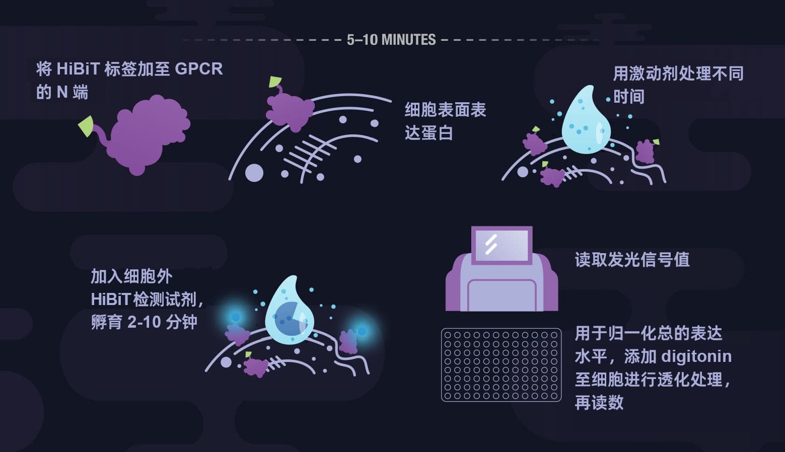
监测受体内化
HiBiT检测方法消除了受体内化研究中基于抗体检测的必要。使用HiBiT技术,操作流程中去除了多个抗体结合步骤和相关的洗涤步骤。只需加入检测试剂,即可测量出发光信号。
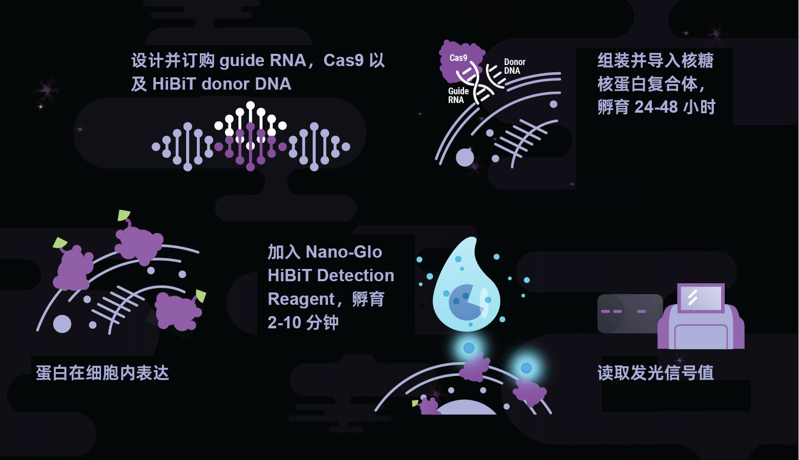
定量蛋白丰度和降解
经典的表位标记方法在通量或灵敏度上是有限的,要求使用高质量的抗体,但也可能只产生半定量的结果。HiBiT标签将生物发光的简便性和灵敏度引入到蛋白质丰度的研究中,可对内源性水平的蛋白质进行定量,即使是那些维持在低表达水平的蛋白质。
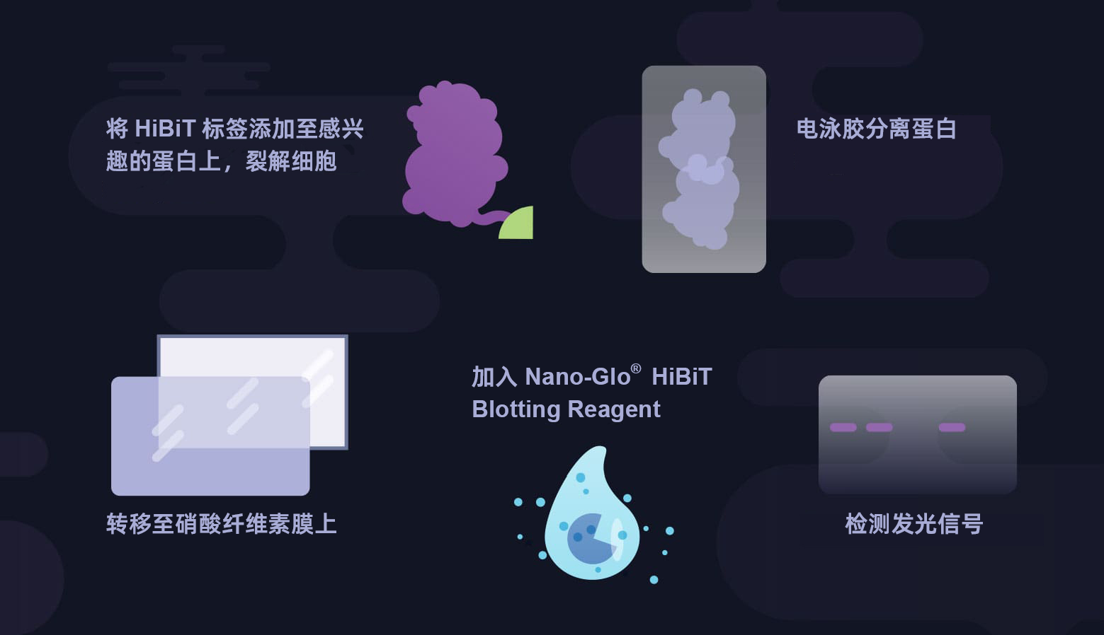
快速简单的免疫印迹检测
1. 什么是HiBiT印迹检测?
- HiBiT印迹检测:一种快速、灵敏的Western Blot替代方法;
- 5分钟内出结果;
- 无需抗体;
- 背景低,灵敏度高;
- 无需洗涤或封闭步骤。
HiBiT是一种基于蛋白质互补的生物发光检测方法。HiBiT标签被添加到你感兴趣的蛋白质中,当互补的LgBiT亚基与HiBiT结合时,形成一个具有功能性的萤光素酶进行检测。
| 2. HiBiT与传统的表位标签不同
| |
| 3. HiBiT蛋白标签的优势
| |
| 4. 如何应用HiBiT标签
| |
参考文献:
1)Nakashoji, A. et al. (2020) Identification of a Modified HOXB9 mRNA in Breast Cancer. Journal of Oncology. Article ID 6065736,
2) Schwinn, M.K. et al. (2020) A Simple and Scalable Strategy for Analysis of Endogenous Protein Dynamics. Sci. Rep. 10(1), 8953.
3) Tange, N. et al. (2020) Staurosporine and venetoclax induce the caspase-dependent proteolysis of MEF2D-fusion proteins and apoptosis in MEF2D-fusion (+) ALL cells. Biomed Pharmacother. 128, 110330.
4) Ranawakage D.C. et al.(2019) HiBiT-qIP, HiBiT-based quantitative immunoprecipitation, facilitates the determination of antibody affinity under immunoprecipitation conditions. Sci. Rep. 9(1) 6895.
5) Sasak, M. et al. (2018) Development of a rapid and quantitative method for the analysis of viral entry and release using a NanoLuc luciferase complementation assay. Virus Res. 243, 69-74.
6) Tamura, T. et al. (2019) In Vivo Dynamics of Reporter Flaviviridae Viruses. J Virol. 93(22), e01191-19.
